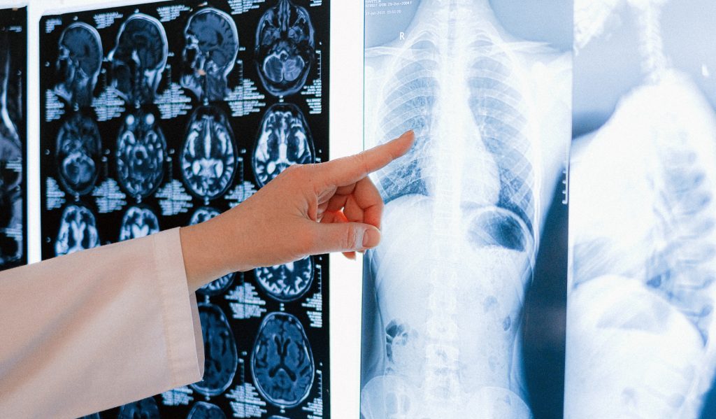
The detection of lung nodules during a CT scan can cause some people to be uneasy and to worry about getting lung cancer. So what is the risk of a lung nodule being cancerous (malignant)? How can we determine it?
Cancer risk of lung nodules
Studies show that over 90% of lung nodules are benign, which may be inflammatory, tuberculosis, benign tumors, etc. On the other hand, only a very small percentage of nodules are cancerous.
Lung nodules are classified according to their density as ground-glass nodules, solid nodules, and part-solid nodules, which are ground-glass nodules with some solid components. Furthermore, there is a correlation between the density of a lung nodule and its risk of cancer. Part-solid nodules have the highest probability of developing cancer, followed by ground-glass nodules and solid nodules.
A cancerous lung nodule usually has certain CT features, such as burrs, lobules, pleural traction, and vesicle signs. Therefore, the risk of a nodule being cancerous is estimated based on its CT features and the patient’s clinical factors, such as smoking history and family history of lung cancer.
High-risk nodules
- Solid pulmonary nodules with a max. size of ≥15 mm, or 8 mm – 15 mm with cancerous CT features (burrs, lobules, pleural traction, vesicle signs, etc.) are high-risk nodules.
- Part-solid nodules with a max. size >8 mm are high-risk nodules.
Management: The larger ones can be diagnosed with a biopsy or other methods and removed by surgery (usually minimally invasive). Others should be followed up to monitor their growth rate at intervals of 1 month, 3 months to 12 months. If nodules are found to grow, surgery should be performed. For those with no change, long-term follow-up is recommended at half the frequency and for a period of not less than 3 years.
Intermediate-risk nodules
- Solid nodules with a max. size of 6 mm – 15 mm and no obvious signs of being cancerous are intermediate-risk nodules.
- Part-solid nodules with a max. size of ≤8 mm are intermediate-risk nodules.
- Pure ground-glass nodules >6 mm in size are considered intermediate risk.
Management: Continuous follow-up of chest CT at 3 months, 6 months, 1 year, and 2 years is recommended. Surgery is recommended for nodules with growth, and long-term CT follow-up of not less than 3 years is recommended for nodules with no change or shrinkage.
Low-risk nodules
- Solid nodules and pure ground-glass nodules with a maximum size of ≤5 mm are low-risk nodules.
Management: CT review is recommended to monitor the growth of the nodule at an interval of 6 months to 1 year for low-risk nodules. Surgery is recommended for nodules with a certain growth rate. Continuation of long-term CT follow-up for no less than 3 years is recommended for nodules with no change or shrinkage.
Conclusion
In conclusion, most lung nodules are benign, and there is no need to be overly concerned. Regular follow-up CT scans are essential to monitor the lung nodules. Once growth is detected, the nodule can be removed surgically (usually minimally invasive), and the prognosis tends to be favorable.
